Cranial Nerves
...
The cranial nerves that can be readily tested are the trigeminal (V), facial (VII), and spinal accessory (XI) (discussed elsewhere).
Facial (VII)
The facial nerve is examined by recording the latency and amplitude from a stimulus at only one site along the course of the nerve. Nerve conduction velocities are not calculated.

Stimulation site: Place the electrodes behind the angle of the jaw, with the cathode posterior to the earlobe and the anode behind. This placement stimulates the nerve just before its enters the parotid gland. Alternatively, you may place the cathode over the stylomastoid foramen and the anode over the mastoid.
Ground: Usually the ground is placed over the parotid area, but you may place it on the chin or forehead also.
Recording sites: Place the active recording over the orbicularis oris at the corner of the mouth, over the orbicularis oculi on the outer canthus of the eye, over the frontalis in the forehead, or over the nasalis muscle on the nasolabial fold. Place the reference electrode on the nose. Either a needle or surface electrode may be used for recording.
The facial nerve may be evaluated differently – by using the blink reflex, which will be discussed with the trigeminal nerve (below).
Trigeminal
This nerve is evaluated by using reflex activity and extrapolating information from it.
Blink reflex
A brief review of the anatomy will assure a better understanding of this study.

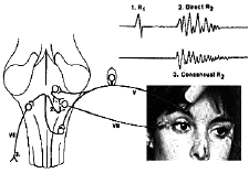
As the sensory fibers of the Vth nerve enter the brain stem, they establish three kinds of synaptic connections with the VIIth nerve nuclei:
– One, a direct and monosynaptic with the ipsilateral VIIth nerve nucleus.
– Another, indirect and polysynaptic with the contralateral VIIth nerve nucleus.
– A third, also polysynaptic, again to the ipsilateral VIIth nerve nucleus.
These connections are demonstrated clinically by the fact that when the glabella is lightly tapped with a reflex hammer or the finger, a brisk blinking reaction is seen bilaterally. The blink reflex is the electrical equivalent of this reaction referred to clinically as the glabellar reflex.
Stimulation of the supraorbital branch of the Vth nerve as it enters the skull through the supraorbital foramen will result in contraction of the orbicularis oculi muscles bilaterally.
The test is best performed by using two channels on the cathode ray tube to study both sides simultaneously.
One each side an active electrode is placed over the orbicularis oculi muscle on the outer canthus of the eye and the reference on the lateral aspect of the nose. One ground is used and is placed over the chin.
The Vth nerve is stimulated via its supraorbital branch over the supraorbital foramen; the sweep speed used is 10 msec/division and the gain set at 200 µv/division. On the ipsilateral channel, both direct and indirect responses are seen, the direct of a short latency and mono or biphasic configuration, the indirect of a long, usually variable, latency and polyphasic configuration. On the contralateral channel, only the indirect, long latency polyphasic response is seen.
Blink Reflex Findings
– In unilateral Vth nerve lesions, all three responses are equally affected.
– In unilateral VIIth nerve lesions, stimulation on the same side of the lesion will result in delayed or absent direct and indirect responses ipsilaterally but a normal indirect response contralaterally. When the nerve is stimulated on the healthy side, both the direct and indirect responses are spared while the contralateral indirect response is affected.
The blink reflex can be used in the evaluation of toxic neuropathies and in comatose patients and multiple sclerosis as a means of evaluating brain stem functions.
Jaw jerk: This response is produced by use of a percussion hammer, which triggers the cathode ray sweep.
Stimulation site: The stimulus is the percussion of the jaw, elicited as in the clinical testing of this reflex.
Ground: The ground may be placed on the chin or the nose.
Recording: Place the active electrodes over the masseter muscles bilaterally and the reference electrodes over the nose or forehead.
The response is bilateral, and it is best to have two active channels on the CRT for comparison.
