Variations In The Innervation Of Upper & Lower Extremities
...
Two relevant variants in the innervation of the upper and lower extremities will be discussed. One is the median-to-ulnar communication, also known as a crossover, and the second is the accessory peroneal nerve.
Crossovers
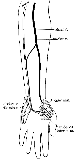
In this instance a few fibers of the median nerve separate from its anterior interosseous branch in the upper third of the forearm, and cross over to join the ulnar nerve.
Crossovers are found in 15 to 30 percent of otherwise normal individuals.

Three types can be seen either separately or in combination:
1 – The hypothenar type, where the crossing fibers end up in the hypothenar muscle group.
2 – The first dorsal interosseous type, where the crossing fibers end up in the first dorsal interosseous muscle.
3 – The thenar type where the crossing fibers go to the ulnar muscles in the thenar group.
In the hypothenar type, routine conductions reveal the following: when the ulnar nerve is stimulated at the wrist, the ADM motor amplitude is much greater than that obtained from stimulation at the elbow (more than 2 mv difference between both responses). This is due to the fact that at the wrist, the nerve contains, in addition to the fibers it already had at the elbow, fibers which have crossed from the median nerve. To differentiate this from an ulnar nerve conduction block at the elbow the following is done: the median nerve is stimulated at the elbow while recording the ADM. If a response is obtained it indicates that there has been a crossover of some median fibers to the ADM since normally, elbow stimulation of the median nerve should not evoke any motor response in the ADM.
In the first dorsal interosseous type, routine conductions do not reveal the cross-over. When the first dorsal interosseous muscle is studied however, the findings are similar to those with the ADM in the hypothenar type. When the ulnar nerve is stimulated at the wrist the first dorsal interosseous motor amplitude is much greater than that obtained when the nerve is stimulated at the elbow (more than the normal 2 mv drop). With median nerve stimulation, the following changes are seen: normally if no crossover is present, a response can be recorded from the first dorsal interosseous on median wrist and elbow stimulation (wrist greater than elbow). When a crossover is present the response from the median elbow stimulation is much greater than that from wrist stimulation because median fibers that will cross to the first dorsal interosseous are still part of the median nerve at this level.
In the thenar presentation, routine studies will reveal the following: when the median nerve is stimulated at the elbow, the APB amplitude is higher than when the nerve is stimulated at the wrist. When the ulnar nerve is stimulated, the APB evoked response is much higher from the wrist than from elbow stimulation. Normally one would obtain a response from the APB from both wrist and elbow stimulation of the ulnar nerve because of volume conduction to the APB from other ulnar innervated muscles in the thenar area. These responses from wrist and elbow stimulation however are usually roughly equal in size. When a crossover is present, the elbow stimulation results in a much lower amplitude than from the wrist because at that level (elbow) the crossover has not taken place yet.
Although median-to-ulnar crossovers do not significantly complicate routine upper extremity workups, they can render the interpretation of nerve conduction studies difficult in the presence of a carpal tunnel or an ulnar neuropathy.
Accessory Peroneal Nerve
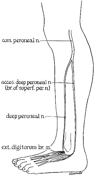
Although the EDB is usually innervated only by the deep peroneal nerve, occasionally (in about one third of the population) it derives additional innervation from an accessory peroneal nerve, a branch of the superficial peroneal, which curves around the lateral malleolus and usually supplies the lateral portion of the EDB.

When the peroneal nerve is stimulated at the ankle, the response obtained has a lower amplitude than that obtained from the knee. This difference is due to the fact that at the ankle only the deep peroneal nerve is stimulated but at the knee both deep and superficial branches (the latter with its accessory peroneal branch) are stimulated.
If the accessory peroneal nerve is stimulated behind the lateral malleolus, a response is obtained from the EDB, with an amplitude that is equal to the difference between the amplitudes of the responses obtained from knee and ankle stimulation.
