The Motor Unit
...
Motor units
In anatomical terms, the motor unit consists of an anterior horn cell, its axon, and all the muscle fibers innervated by that axon and its branches. A motor unit may contain anywhere from a few muscle fibers (in the laryngeal muscle) to several hundreds (in the gastrocnemius).
Muscle fibers belonging to one motor unit are not closely packed together. They are scattered over a small area of muscle and intermingled with fibers belonging to other motor units.
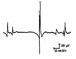
The motor unit action potential is the electrical field generated by muscle fibers belonging to one motor unit as recorded by the tip of the nearby needle electrode.
Normally muscle fibers belonging to one motor unit are all depolarized and repolarized somewhat synchronously.
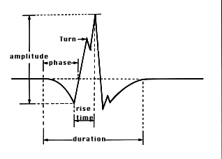
Amplitude, duration, number of phases, rise time, and firing rates characterize a motor unit potential. Traditionally one measures the amplitude from peak to peak; the duration from the first deflection of the baseline to the last return to it; the number of phases by counting the number of times the components of the motor unit potential cross the baseline plus one; and the rise time as that elapsed between the peak of the initial positive (down) deflection to the peak of the highest negative (up) deflection.
Note, however, that the number of fibers contained in a motor unit and their degree of synchrony affect those characteristics.
The number of phases a motor unit contains depends largely on the synchrony of depolarization of its muscle fibers and can be affected either by nerve disease causing differential slowing in impulse conduction, or muscle disease where the conduction characteristics of the muscle fibers themselves have changed.
The rise time, strictly a function of the proximity of the needle tip to the muscle fibers of the contracting unit, is usually between 200 and 300 µsec.
The firing rates of motor units depend on their type and size. Smaller units are recruited early, with weak effort, and fire faster than large units which are recruited later as effort is increased.
All the above characteristics vary with age, with the muscle under study, and with muscle temperature. Minute changes in needle position can greatly affect the shape of the motor unit potential. At a distance of 0.12 mm of the depolarized fibers, the amplitude may be decreased by as much as 50 percent and at l mm by an astounding 90 percent.
In view of these variations, when single estimates of size and duration from quick “eyeballing” of motor units is a problem, reading should be done either by storing samples of the unit, by photographing the unit or, better still, by having the unit trigger the sweep and using a delay line to permit their study in detail.
Temperature Effect: At lower temperatures the motor unit duration and its amplitude are increased.
NEEDLE EXAM DESCRIPTION
There are four stages in the examination of a muscle by needle electrode: when the muscle is at rest and during mild, moderate and full voluntary effort.
The Muscle At Rest
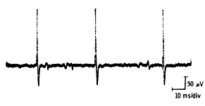
Insertional activity: The response of the muscle fibers to needle electrode insertion is called the insertional activity. Normally it consists of brief, transient muscle action potentials in the form of spikes, lasting only a few seconds and stopping immediately when needle movements stop. Note that insertional activity may be decreased, such as in fibrosis or fat tissue replacement; or prolonged, such as in early denervation (the so-called irritability) and in myotonic disorders.
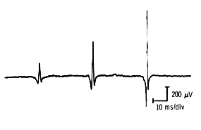
Spontaneous activity: The persistence of any activity beyond insertion constitutes spontaneous activity. This could be due to the normal end-plate noise, or to the presence of fibrillations and positive waves, or other spontaneous activity (see below).
A normal spontaneous activity is the end-plate noise. This can either be monophasic (end-plate noise) or biphasic (end-plate spikes) potentials, recorded when the needle is in the vicinity of a motor end-plate.
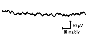
The monophasic potentials are of low amplitude and short duration and cause a “thickened baseline” appearance. They give a typical “sea shell” noise or “roar” on the loudspeaker.
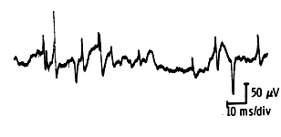
The biphasic activity consists of irregular biphasic, 100-300 µv spikes of short duration.
The muscle at rest must be examined in four or five different directions once the needle is inserted to ensure adequate sampling. A pause of 0.5-1 second is required between each insertion to allow for the observation of any spontaneous activity. When fasciculations are suspected, this time is less than adequate and a 10 to 15 second pause is more appropriate.
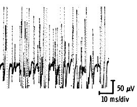
For optimal observations of insertional activity set the oscilloscope sweep speeds at 10 ms/division and amplification at 50 – 100 µv/division. Filter settings chosen are 32 Hz for the low frequencies and 8000 or more Hz for the high.
The Muscle During Voluntary Effort
Assess voluntary activity during three stages of effort: mild, moderate, and full. With mild and moderate voluntary effort, individual motor units can be studied separately and their amplitude, duration, and number of phases measured. Recruitment and firing rates are best assessed during moderate effort, the interference pattern during full effort.
Mild effort: Only a few motor unites are observed at this stage. These are the smaller motor units as they are the ones to be recruited first. Ask the subject to maintain a steady minimal contraction and sample the muscle in four or five different areas. Sample at least 20 motor units and calculate an average amplitude, duration and number of phases.
Moderate effort: The firing rates and recruitment of motor units are best assessed during this stage. As muscle effort increases, motor unit firing rates are increased and new motor units are recruited. The units seen at this stage are larger than those seen with mild effort.
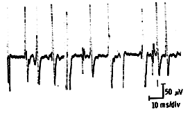
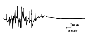
Full effort: At maximum contraction, the firing rates go even higher and more motor units are recruited into the contraction making it difficult to distinguish them individually. When all the motor units are recruited a complete interference pattern is observed.
Motor unit potentials are best studied with the same filter setting used for insertional and spontaneous activity, i.e. 16-32 Hz low and 8000 Hz or more high. Motor unit potentials’ duration is measured with an amplification setting of 100-200 µv/division, and their amplitude at settings of 500 µv – 2000 µv/division, depending on the size of the motor unit under study. The sweep speed setting is 5-10 ms/division. While these settings are fairly widely accepted, different labs use different individual settings. It is essential however to use the same settings consistently to perform motor unit potential measurements.