Pathological Processes
Pathological processes that affect nerve conduction studies
Knowing how different pathological processes affect nerve conduction studies underlies the understanding and interpretation of nerve conduction findings. The following changes will be discussed:
Demyelination
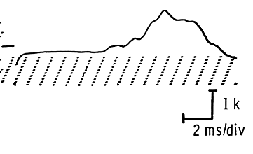
As a rule, latencies and conduction velocities are affected most. With few exceptions, the sensory fibers are affected first. The sensory nerve action potential’s duration is increased, resulting in a low amplitude and prolonged distal latency. At a later stage, the motor fibers are affected essentially in the same fashion with decreased conduction velocities, usually 50 percent below normal values.
In advanced demyelination, sensory responses may be altogether absent.
In the entrapment or pressure neuropathies, the demyelination is focal, with the nerve remaining normal both above and below the lesion. When the nerve is stimulated above the entrapment or pressure area, the conduction velocity is slowed. Stimulation below the lesion, however, results in a normal velocity.
In distal entrapments, where stimulation below the lesion is either impossible or technically difficult, the findings are limited to a prolonged distal latency and a reduction of the sensory amplitude and, in time, of the motor responseTemporal dispersion

In polyneuropathies, the above changes are present diffusely, although they may be more severe at or about potential pressure and entrapment points.
Conduction Block
The cause of conduction blocks is unclear. Such blocks can arise from a severe focal demyelinating lesion, making impulse propagation through the area of demyelination impossible; or from physiological interruption of conduction without detectable abnormalities histologically. Below the lesion, the nerve conducts the impulse normally.
A partial block is one in which only a few fibers are affected. The nerve can still be stimulated above the lesion, but, since only a few fibers conduct, a low amplitude response is obtained.
When the block is complete, no response can be obtained on stimulation above the lesion. When stimulation below the lesion is possible, a normal response is seen.
Sometimes partial conduction blocks are seen along with focal demyelinating lesions. Stimulation above the lesion yields a low amplitude response with a slowed conduction velocity along the involved segment. Stimulation below the lesion, when feasible, results in a normal amplitude and conduction velocity.
Axonal Loss
In contrast to the effects of myelin-sheath lesions, loss of axons results primarily in decreased amplitudes. The sensory fibers are affected first with a resulting decreased in amplitudes but relatively preserved distal latencies. As the lesion becomes more severe, motor amplitudes are decreased and sensory potentials may even become unobtaibnable.
In advanced disease, the motor amplitudes may be so depressed that motor distal latencies are prolonged and conduction velocities slowed, though usually the slowing does not fall below 30 percent of the expected normal value. These effects result from a dropout of the fastest conducting fibers.
In contrast to amplitude effects in lesions with conduction block, low amplitude from axonal loss cannot be corrected by stimulation below the lesions. In conduction blocks, the nerve segment below the lesion is normal, whereas in axonal loss it undergoes Wallerian degeneration and does not conduct normally distal to the lesion. An evoked response can still be obtained however, from stimulation below the level of the injury, up to ninety-six hours after total nerve transection.
